Two names appear repeatedly in research on Chinese skullcap: baicalein and baicalin. Chemically, they differ by remarkably little. Baicalein is the bare flavone; baicalin is baicalein with a glucuronic-acid group attached at one position.
That single addition changes far more than the name. It changes how the molecule crosses the gut, what the liver does to it, which forms dominate after absorption, and — in some cancer models — even the biological route by which the two compounds produce an effect.
There is one complication: the body converts them back and forth.12 So baicalein and baicalin are neither completely separate drugs nor interchangeable versions of the same one. They are better understood as two members of a continuously interconverting metabolic pair.
One sugar changes the journey
Both compounds originate from the root of Scutellaria baicalensis. Baicalein is 5,6,7-trihydroxyflavone — the aglycone, or unconjugated flavone. Baicalin is baicalein 7-O-glucuronide: the same flavone scaffold carrying a glucuronic-acid group.
In the plant, baicalin is the major stored form.3 But the two compounds face quite different problems once swallowed.
Baicalein is relatively membrane-permeable and is absorbed quickly, but it is poorly soluble and undergoes extensive first-pass metabolism. The intestine and liver rapidly attach glucuronide and sulfate groups to it, so only a small part of the circulating pool remains as free baicalein.45
Baicalin starts from the opposite problem. Despite being more polar, it has low aqueous solubility and low intestinal permeability and is poorly absorbed intact.6 Much of an oral dose first has to be stripped of its glucuronide by bacterial β-glucuronidase in the gut, regenerating baicalein. That aglycone crosses the intestinal wall much more readily — after which intestinal and hepatic enzymes promptly conjugate much of it again.1
Same flavone scaffold. Two quite different routes into the body.
Your gut and liver keep rewriting the label
The conversion is not simply baicalin → baicalein.
Gut bacteria hydrolyse baicalin into baicalein. The absorbed baicalein is then glucuronidated again — particularly through UGT enzymes such as UGT1A9 — while sulfated and other glucuronidated metabolites are also formed.5 Conjugated material can be pumped into bile or back toward the intestine, where bacterial enzymes can remove the glucuronide again.17
That recycling helps produce the characteristic multiple peaks seen in pharmacokinetic studies.1
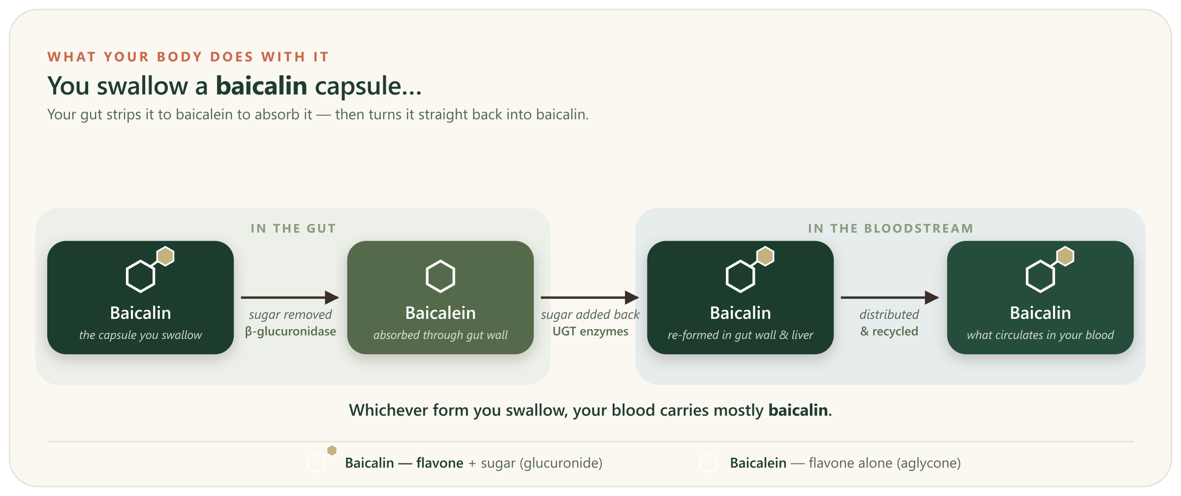
Starting with baicalein does not escape the cycle either. Human studies show rapid conjugation after absorption, with circulating sulfate and glucuronide metabolites reaching considerably higher concentrations than the unconjugated parent compound.8
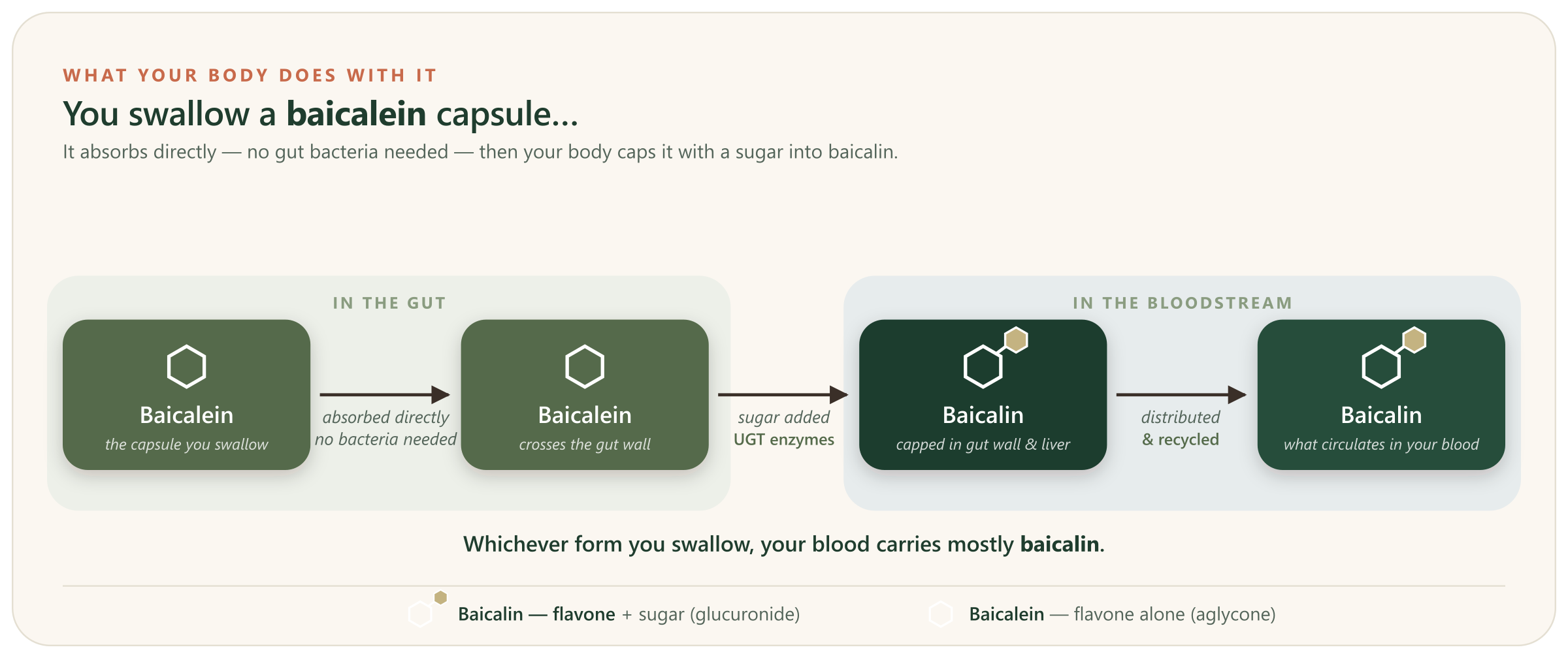
So the useful mental model is not baicalein or baicalin, but baicalein ⇆ baicalin + other conjugated metabolites. The name on the bottle tells you the compound entering the system. It does not fully describe the chemical mixture circulating later.
The two, side by side
The table below exposes something more interesting than “one has a sugar.” The two compounds share a great deal of cancer biology — but they do not always arrive at it by the same route.
| Property | Baicalein | Baicalin |
|---|---|---|
| Chemical form | Aglycone — the unconjugated flavone. | Baicalein 7-O-glucuronide. |
| Main absorption problem | Rapidly absorbed, but limited by poor solubility and extensive first-pass conjugation.45 | Low solubility and low permeability; much oral absorption depends on bacterial conversion to baicalein.61 |
| Human PK evidence | Several Phase I studies; parent and metabolite concentrations measured.891011 | Sparse direct human PK; its own absolute oral bioavailability and Cmax have not been established.9 |
| Distinctive glycolysis mechanism | Suppresses HIF-1α-driven glycolysis in several tumour models.121314 | Promotes degradation of hexokinase-2 in a colorectal-cancer model, with downstream cGAS/STING immune signalling.15 |
| Distinctive cell-state finding | Drove apoptosis in a direct colon-cancer comparison.16 | Drove cellular senescence in the same model.16 |
| Ferroptosis | Predominantly ferroptosis-inhibiting in the lab, although pro-ferroptotic exceptions exist.1718 | Repeatedly ferroptosis-inducing in tumour models, although it can protect normal tissue from oxidative death.1920212223 |
| Angiogenesis | Relatively coherent anti-angiogenic evidence, including TLR4→HIF-1α→VEGF suppression.24 | Mixed: anti-angiogenic tumour findings coexist with an independent pro-angiogenic VEGF result.2526 |
| Human oncology evidence | None. | None. |
| Interaction emphasis | Antiplatelet activity; CYP3A4 / P-gp signals, mostly preclinical.2728 | Antiplatelet activity plus a documented human pharmacokinetic interaction with rosuvastatin.2930 |
Most of their biology actually overlaps
It would be easy to turn the baicalein–baicalin story into a neat tale in which attaching a sugar creates a completely different compound. The research does not support anything that simple.
Both have been reported to suppress major cancer-associated signalling networks including STAT3, PI3K–AKT–mTOR, ERK and Wnt/β-catenin; both can reverse epithelial–mesenchymal transition; both can trigger mitochondrial apoptosis; and both have suppressed tumour growth in animal models.313
The difference is therefore not that one molecule is “anticancer” while the other behaves differently. It is subtler: the glucuronide changes exposure, cellular access and the relative importance of particular mechanisms. That is precisely why apparently small chemical modifications matter so much in pharmacology.
Where the difference becomes genuinely striking
The cleanest example comes from a study that tested the two compounds in the same colon-cancer system. Baicalein pushed the cells predominantly toward apoptosis — controlled cellular self-destruction. Baicalin instead produced senescence, a durable growth-arrest state, including suppression of telomerase activity.16 A separate baicalin study later reproduced a senescence programme through DEPP, Ras/ERK and p16/Rb signalling.32
The difference becomes even more pronounced with ferroptosis, an iron-dependent form of oxidative cell death. Baicalein is well known as a lipoxygenase inhibitor and lipid-radical scavenger, properties that can interrupt the lipid oxidation needed for ferroptosis; accordingly, much of its ferroptosis literature is protective.17 But that direction is not absolute: in particular tumour settings baicalein can indirectly push cells into ferroptosis by suppressing upstream survival signalling and GPX4.18
Baicalin’s tumour literature points much more consistently the other way. Ferroptosis induction has been reported through several different mechanisms, including suppression of the xCT/GPX4 defence system, regulation of ALOX12, p53 signalling and mitochondrial iron handling.19202122 So “baicalein blocks ferroptosis and baicalin causes it” is too simple — but the balance of the evidence really is different.
Angiogenesis tells a similar story. Baicalein has a comparatively coherent anti-angiogenic record, including suppression of TLR4/HIF-1α/VEGF signalling in a colorectal tumour model.24 Baicalin also has anti-angiogenic tumour data — but an independent study found it increasing VEGF and angiogenesis through ERRα/PGC-1α.2526 That contradiction is useful: it is a reminder that conjugation can alter biological context rather than merely weakening or strengthening the same effect.
They even attack the same metabolic problem differently
Both compounds have been linked to suppression of the Warburg phenotype — cancer’s heavy reliance on glycolysis — but the strongest mechanisms are quite different.
For baicalein, the recurring node is HIF-1α. In breast, oesophageal and gastric cancer models it reduced HIF-1α-dependent glycolytic signalling, lowering glucose use, lactate production or glycolytic enzymes and, in some models, restoring sensitivity to therapy.121314
Baicalin has a newer and unusually direct mechanism. In a colorectal model it promoted proteasomal degradation of hexokinase-2, an important glycolytic enzyme. The resulting mitochondrial damage released mitochondrial DNA, activating cGAS/STING signalling and shifting tumour-associated macrophages toward a more inflammatory, tumour-opposing phenotype.15
Both findings remain preclinical. But mechanistically they are not duplicates: one form acted on a transcriptional controller of tumour metabolism; the other targeted a glycolytic enzyme and connected metabolic disruption to the tumour immune environment.
Why the starting form still matters
If the body converts one into the other, it is reasonable to ask why a baicalein supplement should be distinguished from a baicalin supplement at all. Three reasons.
The first is exposure. Baicalein crosses the intestine relatively readily and is then heavily metabolised; baicalin is poorly absorbed intact and depends much more strongly on gut-bacterial hydrolysis before absorption.46 Starting with different forms therefore changes the route, timing and likely local concentrations encountered before the molecules enter the same metabolic cycle.
The second is evidence. Baicalein has been studied directly in several Phase I pharmacokinetic trials; at a repeated human dose of 1,800 mg/day, measured parent baicalein reached a steady-state peak of roughly 4.9 µM, while conjugated metabolites circulated at considerably higher concentrations.8 Direct human data for purified baicalin are much thinner — small pharmacology studies exist, but its own absolute oral bioavailability and peak plasma concentration have not been established.91 So the confidence with which we can describe human exposure differs even though the two molecules interconvert.
The third is drug interactions. Their profiles overlap but are not identical. Both carry preclinical antiplatelet signals. Baicalein also has CYP3A4 and P-glycoprotein interaction evidence.2728 For baicalin, a human study found a substantial genotype-dependent reduction in rosuvastatin exposure during repeated dosing — one of the rare direct human interaction signals in this literature.2930 The starting compound therefore still matters clinically even when metabolism later blurs the boundary.
The number that keeps the laboratory results in perspective
One final difference is worth keeping in view, because it changes how the cancer research should be read.
For baicalein, we actually have a human benchmark. At 1,800 mg/day in a pharmacokinetic study, parent baicalein reached a peak of about 4.9 µM.8 Many of the cell experiments reporting apoptosis, pathway suppression or other anti-tumour effects used roughly 25–100 µM.33 Some molecular effects occur closer to measured human exposure — the colorectal ferroptosis work was active from about 7.5 µM — so the gap is not universal, but it is substantial for much of the cell literature.18
For baicalin the uncertainty is greater. Many mechanistic cancer studies use roughly 10–200 µM, while no human study has established the peak concentration reached after a purified oral baicalin dose.34359 Animal pharmacokinetic work suggests low intact exposure, yet animal tumour studies show that oral dosing can nevertheless produce biological effects despite extensive conversion and recycling.15 That is why the animal studies matter — and why they still cannot answer the human question.
Neither compound has demonstrated anti-cancer efficacy in people.
Where this leaves things
Baicalein and baicalin are close enough chemically that the body repeatedly converts one into the other, but different enough pharmacologically that treating them as interchangeable loses important information. The glucuronide changes the route of absorption. It changes the balance of circulating forms. It changes which interactions have actually been measured. And in experimental cancer biology it sometimes changes the mechanism itself — apoptosis versus senescence, predominantly anti- versus pro-ferroptotic behaviour, clean versus contradictory angiogenesis signals, HIF-1α versus hexokinase-2-driven metabolic effects.
That is the useful lesson of the pair. A sugar attached to a flavone is not decorative chemistry. It can change where the molecule goes, what cells see, and what the evidence actually means. None of it is a cancer-treatment claim — but if you take skullcap in any form, tell your care team, because the gap between a dish and a human body is exactly where these distinctions live.
← All articles