Cancer is complicated. That statement is so familiar it risks becoming meaningless. But one particular kind of that complexity matters enormously when we talk about cancer pathways: a cancer’s aggressiveness is not the same thing as its adaptability. Holding those two ideas apart is the key to reading any pathway map — including the Mechanica Natura Pathway Atlas — for what it can and cannot tell us.
An aggressive cancer may grow rapidly, invade early, or become immediately life-threatening while remaining unusually dependent on a narrow molecular program. Canonical PML::RARA acute promyelocytic leukemia (APL) is the classic example. Historically one of the most dangerous acute leukemias, its malignant identity is dominated by a single fusion protein, PML::RARA. Drugs that directly dismantle that program, principally all-trans retinoic acid and arsenic trioxide, have turned APL into one of oncology’s great examples of a molecularly targeted cure.[1]
It is worth being precise about what that example does and does not show. Aggressiveness and adaptability often travel together, because genomic instability and intratumour heterogeneity enlarge the pool of variants that treatment can then select from. But adaptability cannot be inferred from proliferation rate or genomic instability alone, and it can arise with no new mutation at all. APL is striking precisely because its routes of escape are comparatively narrow. It is not beyond adaptation — under sustained pressure, resistance can still emerge — but its survival rests on an unusually narrow breadth of dependency, which is exactly what makes it so treatable.
At the other extreme are cancers with extraordinary biological flexibility. Inhibit one signalling route and another becomes dominant. Restrict one nutrient and another fuel is scavenged. Eliminate one cell population and a resistant one expands. In some cases, cancer cells can even change their transcriptional or lineage identity.
What “adaptability” means in this article
Because the word is doing a lot of work here, it helps to be explicit. “Adaptability” is not a single, standardized clinical measurement the way tumour grade, stage, or proliferation rate is. In this article it is a plain-English umbrella for several genuinely distinct ways a cancer can preserve fitness when its current state is disturbed:[2]
- Rapid network feedback (minutes to hours): blocking one node releases a brake elsewhere.
- Reversible non-genetic states: transcriptional, epigenetic, or metabolic changes, including drug-tolerant “persister” states, that require no new mutation.
- Clonal selection: expansion of resistant subclones already present at low frequency before the perturbation.
- Genetic evolution: acquisition, then selection, of new resistance-conferring alterations during continued treatment.
- Lineage or cell-state transformation: the tumour changes the very identity on which the original treatment depended.
Modern reviews explicitly separate these non-genetic adaptive processes from genetic evolutionary routes to resistance, and note that the two are often linked.[2] Keeping them distinct matters, because the examples later in this article each illustrate a different one, and a pathway map treats them very differently.
First problem: there is no definitive number of human pathways
At the time of writing, the Mechanica Natura Pathway Atlas tracks 74 cancer-relevant metabolic and signalling pathways, and that number grows as the evidence base does. It is tempting to imagine that somewhere beyond them lies a definitive master list, perhaps hundreds or thousands of pathways that, if catalogued completely, would finally describe the system.
Biology does not divide itself so neatly.
A biochemical or signalling “pathway” is a model imposed on an interconnected network. Researchers draw boundaries around a sequence of reactions or signalling events because doing so makes the system understandable and experimentally tractable. But proteins, metabolites, and regulatory molecules do not necessarily respect those boundaries. A single receptor may activate several downstream networks. AKT participates in metabolism, growth, apoptosis, protein synthesis, and stress responses. mTOR integrates growth-factor signalling with amino-acid, energy, and oxygen availability. 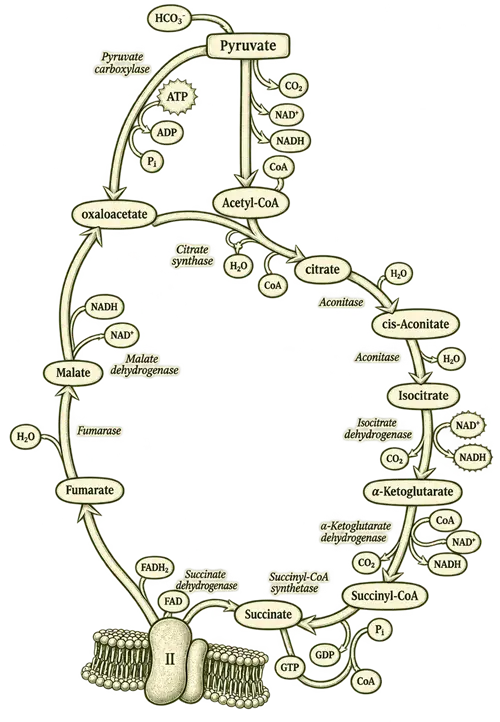 The citric acid (Krebs) cycle. Only one metabolic pathway but already intricate.MYC drives the cell-cycle machinery while simultaneously reshaping glucose, glutamine, nucleotide, and lipid metabolism. Reactive oxygen species can be metabolic by-products, signalling molecules, and sources of cellular damage all at once.
Even the granularity of a “pathway” is negotiable. PI3K–AKT–mTOR can reasonably be shown as one pathway. It can equally be split into PI3K signalling, AKT signalling, mTORC1, mTORC2, AMPK–mTOR regulation, insulin/IGF signalling, amino-acid sensing, and multiple downstream branches. RTK–RAS signalling can be drawn as one canonical pathway or separated into EGFR, HER2, MET, FGFR, the RAS–RAF–MEK–ERK cascade, and numerous feedback circuits.
This is why the honest answer is that there is no universal, ontology-independent denominator. Curated databases such as Reactome or KEGG do publish pathway counts, and within any one of them a count is perfectly meaningful. But those counts are products of that database’s chosen granularity, not a reading of some objectively correct number of “pathways in a human” or “pathways in cancer.” No such number exists. So there is no scientifically meaningful sense in which the atlas covers “70-plus out of X total cancer pathways.”
Douglas Hanahan’s evolving Hallmarks of Cancer framework makes much the same point at a higher level. The hallmarks are explicitly a heuristic, a way of reducing extraordinary biological diversity into useful organising principles. As knowledge expanded, phenotypic plasticity, non-mutational epigenetic reprogramming, senescent cells, and polymorphic microbiomes all had to be folded into the model.[3] A good framework becomes more useful by organising complexity. It does not make the underlying complexity disappear.

Two maps that illustrate the problem
Stanford Medicine, through its Department of Biochemistry, has produced a remarkable visual resource, the Stanford Pathways of Human Metabolism map. It integrates metabolites, enzymes, cofactors, and major metabolic processes into a single interconnected diagram used in Stanford’s medical biochemistry teaching. Stanford itself is careful to qualify it. The map is “a comprehensive overview of human metabolism,” but, in its own words, “while not exhaustive, the content was selected to illustrate key metabolic pathways and their interrelationships.”[4]
→ Request the Stanford Pathways of Human Metabolism map (free): metabolicpathways.stanford.edu ↗
The scale of that map is instructive precisely because it covers metabolism alone. Add growth-factor signalling, transcriptional regulation, cell-cycle control, apoptosis, DNA repair, chromatin regulation, immune signalling, extracellular-matrix interactions, stress responses, and cell-to-cell communication, and the network expands dramatically.
The TCGA PanCancer Atlas approaches the problem from the opposite direction. In one landmark analysis, investigators examined 9,125 human tumours across 33 cancer types and 64 tumour subtypes, but deliberately organised the analysis around just 10 canonical oncogenic signalling pathways: cell cycle, Hippo, MYC, Notch, NRF2, PI3K–AKT, RTK–RAS, TGF-β, p53, and WNT/β-catenin. Eighty-nine percent of the tumours carried at least one driver alteration within those ten pathways.[5] But it does not mean the pathway governed the tumour, and it does not mean that reversing the alteration would undo the cancer. The value of the study never depended on pretending cancer contains only ten pathways. The investigators reduced a vastly larger network to a set of recurrent organising systems that could be compared systematically across cancers. The distance between “carries an alteration here” and “depends on this” is the whole subject of the rest of this article.
→ Explore the TCGA PanCancer Atlas interactive resource: cell.com PanCancer Atlas ↗
That is essentially what every pathway atlas does. It chooses a level of resolution at which biology becomes navigable.
But the networks do not merely overlap. They respond.
The harder problem is that a cancer pathway diagram is closer to a snapshot than to a permanent wiring diagram.
Cancer cells live inside regulatory systems full of positive feedback, negative feedback, redundant routes, and compensatory mechanisms. Some adaptations unfold over months or years through the selection of resistant cells. Others appear within hours, because blocking one node removes a feedback brake that had been quietly suppressing another.
It is worth correcting a common oversimplification here. Adaptation does not always wait for a brand-new mutation to appear. A resistance-conferring subclone can already exist at very low frequency before treatment starts and simply expand once therapy clears the field around it. A cell can also enter a reversible, drug-tolerant state that requires no mutation at all, survive the initial assault, and only later acquire stable genetic resistance. In experimental models of EGFR-mutant lung cancer, the same T790M resistance mutation could arise by two distinct routes: selection of pre-existing T790M-positive cells, or later genetic evolution of initially T790M-negative drug-tolerant cells; cultures derived from resistant patient tumours supported the clinical relevance of both.[6] Selection, reversible non-genetic states, and genetic evolution are different trajectories that can even occur in sequence.[2]
This is why an arrow on a pathway diagram can mislead when read too literally. If inhibiting pathway A automatically activates pathway B, then calling a compound “an inhibitor of pathway A” tells us very little about the eventual state of the tumour. We already have vivid experimental examples of exactly this.
Six ways the map moves
The examples above are not a random gallery. Each illustrates a different kind of adaptation, and together they trace several of the ways a static diagram can go quietly out of date.
| Type of adaptation | What happens | Example in this article |
|---|---|---|
| Rapid network feedback | Blocking one node releases a brake on another within hours. | KRAS or BRAF blockade reactivates EGFR; catalytic mTOR inhibition restarts AKT. |
| Metabolic reallocation | Impairing one fuel system raises dependence on another. | Pancreatic cancer: ERK inhibition drives up autophagy. |
| Bypass and cell-state change | A parallel receptor, or a new lineage, sustains survival. | EGFR-lung: MET bypass, then small-cell transformation. |
| Reversible oncogene dosage | The tumour lowers, then later restores, how much of the targeted oncogene it carries. | Glioblastoma: mutant EGFR on extrachromosomal DNA. |
| Lineage plasticity | The cancer abandons the pathway’s cell identity entirely. | Prostate: progression to AR-independent, neuroendocrine-like disease. |
| Signals from outside the cell | A neighbouring cell supplies the escape signal. | Melanoma: stromal HGF activates tumour-cell MET. |
A pathway is therefore not the same thing as a vulnerability
This distinction matters most when interpreting nutraceutical research. Showing that a compound affects NF-κB, PI3K–AKT–mTOR, WNT/β-catenin, STAT3, autophagy, or glycolysis establishes a biological interaction. It does not establish that the affected pathway is a dominant vulnerability in every cancer, or even in every tumour carrying the same mutation. Several further questions follow immediately:
- Is the cancer actually dependent on that pathway?
- Does the compound reach it at concentrations achievable in humans?
- Is the direction of effect consistent across tumour contexts?
- What parallel pathways remain available?
- Does suppressing the pathway release a feedback loop?
- Does the tumour switch fuels or ramp up autophagy?
- Can another subclone expand?
- Does the microenvironment supply the missing signal or metabolite?
- And does prolonged pressure eventually select a different cellular state?
There is also a quieter point here. Nearly all of the examples above come from potent, targeted anticancer drugs, so the same adaptive responses cannot simply be assumed for a nutraceutical that modulates the same pathway. Whether a compound engages a target enough to provoke a compensatory response depends on the compound, the achievable exposure, the pathway, and the tumour context. Weaker modulation may apply less selective pressure — but it may equally be too weak to produce a meaningful antitumour effect in the first place. Neither therapeutic activity nor adaptive escape can be read off a pathway count. This is part of why these compounds tend to sit alongside cancer care rather than stand in for it, and why the molecules are often at their most powerful not in the bottle but as a starting point: isolated from the plant, chemically refined, and given at exposures no oral (retail) supplement can reach, several plant natural products have been developed into approved cancer drugs, a path traced in From Folk Remedy to Chemotherapy.
Even genuine multi-pathway activity does not settle the matter. Hitting five nodes weakly is not automatically better than hitting one indispensable dependency hard. And the reverse can also be true: modulating several genuinely complementary vulnerabilities at once may behave very differently from touching each alone. Pathway count is not therapeutic potency.
Why “just” over 70 pathways?
The Mechanica Natura Pathway Atlas is not meant to describe every biochemical reaction, signalling event, or adaptive state that can occur in every cancer. It could not. Its pathways are a curated set of recurrent, cancer-relevant mechanisms for which meaningful nutraceutical-interaction research exists. Some are large signalling systems. Some are narrower metabolic dependencies. Some overlap deliberately, because they capture biologically useful distinctions at different levels of resolution.
They should be read as coordinates, not compartments.
A compound appearing against several pathways means published research has connected it to those biological processes. A cancer associated with one of those pathways means research supports a role for that mechanism in at least some relevant disease contexts. Neither statement means the pathway acts in isolation, that it is equally important in every tumour, or that altering it will necessarily produce a therapeutic effect. The atlas is a map of evidence, not a model that can predict an individual cancer.
The map will never equal the territory
Cancer biology is being resolved at extraordinary speed. Genomic sequencing identifies mutations and copy-number changes. Transcriptomics reveals which genes are actually expressed. Proteomics and phosphoproteomics show signalling activity. Metabolomics and isotope tracing follow nutrient use. Single-cell methods expose the different populations sharing one tumour. Spatial methods show where those populations sit, and help reveal how they interact with the stromal and immune cells around them.
Each layer makes the map better. Each layer also reveals more complexity. That is not a failure of cancer research. It is what increasingly accurate science looks like.
The Stanford metabolic map is enormous and openly not exhaustive. The PanCancer Atlas transformed our understanding of oncogenic signalling while intentionally concentrating on ten canonical pathways. The Mechanica Natura atlas faces the same underlying abstraction problem, at a very different scale and for a very different purpose: select biologically important systems, organise the evidence around them, and make their relationships easier to understand.
Over seventy pathways is therefore both a great many and nowhere near enough to contain cancer biology. Adding the next pathway will not complete the picture. Neither would a hundred more.
There are pathways not represented here. There are interactions no one has characterised yet. There are tumour-specific dependencies that vanish under treatment and new ones that emerge. There are metabolic adaptations that shift with nutrient availability, metastatic site, and microenvironment. There are compounds whose actions remain incompletely understood, and cancers whose relevant vulnerabilities change over the course of their own evolution.
No website can answer for every pathway × every compound × every cancer × every mutation × every disease stage × every treatment context. Current science cannot comprehensively answer all of those combinations either. That limitation should not make pathway research less useful. It tells us how to use it correctly.
A pathway atlas is not a blueprint of cancer. It is a navigation system through what we currently know. And like every good map, its value comes not from pretending to contain the whole territory, but from being clear about what it shows, what it leaves out, and where the edges of present knowledge begin.